International Conference on Medical Image Computing and Computer Assisted Intervention (MICCAI), Oct. 2020.
Automated Measurement of Key Morphological
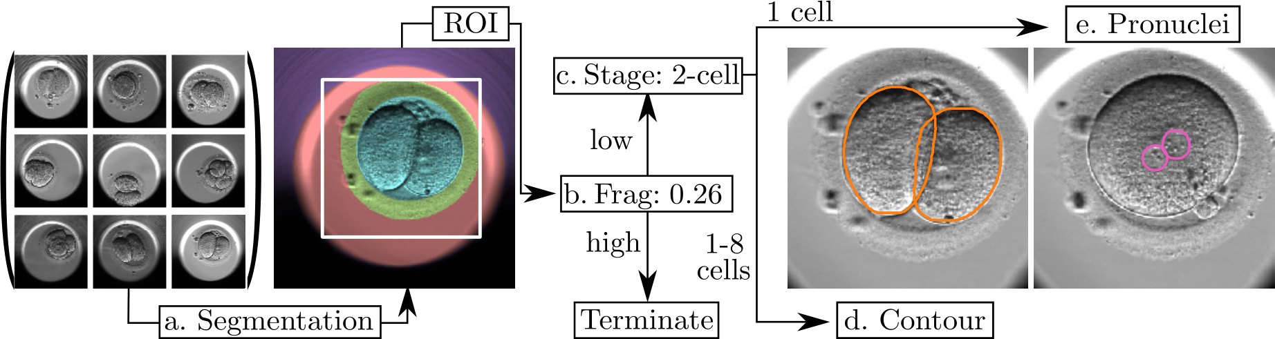
Features of Human Embryos for IVF
Features of Human Embryos for IVF
Authors
Helen Y. Yang*
Harvard University
Harvard University
Robbert Struyven
Harvard University
Harvard University
Zhe Sun
Harvard University
Harvard University
Kylie R. Lee
Harvard University
Harvard University
Charlotte Royston
Harvard University
Harvard University
Liz Cam
Harvard University
Harvard University
Yael Kalma
Tel Aviv Sourasky Medical Center
Tel Aviv Sourasky Medical Center
Foad Azem
Tel Aviv Sourasky Medical Center
Tel Aviv Sourasky Medical Center
Dalit Ben-Yosef
Tel Aviv Sourasky Medical Center
Tel Aviv Sourasky Medical Center
*These authors contributed equally to this work
Abstract
Paper
arXivSource code
GitHub repoExperiental results
Citation
@inproceedings{leahy2020automated,
title={Automated Measurements of Key Morphological Features of Human Embryos for IVF},
author={Leahy, BD and Jang, W-D and Yang, HY and Struyven, R and Wei, D and Sun, Z and Lee, KR and Royston, C and Cam, L and Kalma, Y and Azem, F and Ben-Yosef, D and Pfister, H and Needleman, D},
booktitle={MICCAI},
year={2020}
}